tree in bud opacities pneumonia
Visible thickened visceral and parietal pleura with fluid collection in between Suggests the presence of empyema. It occurs in acute tuberculosis but also in any other bacterial infection.
2
Extensive cavitation of the tuberculous pneumonia lead to endobronchial spread of the infection.

. Additionally some miliary opacities are very dense narrowing the differential - see multiple small hyperdense pulmonary nodules. Tree in bud pattern mosaic attenuation and mucus impaction. Tree in bud opacity.
Causes and imaging patterns of tree-in-bud opacities. Rupture of necrotic lymph nodes into the bronchi can also result in endobronchial dissemination. Tree-in-bud describes the appearance of an irregular and often nodular branching structure most easily identified in the lung periphery.
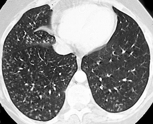
Crossref Medline Google Scholar. Classically bronchiolitis appears as a region of centrilobular nodularity often in a tree-in-bud pattern. Visible small airways or terminal bronchioles filled with mucus pus fluid or cells forming impactions that resemble a budding tree with branching nodular V and Y shaped opacities Split pleura sign.
Bat wing opacities also known as butterfly opacities refer to a pattern of bilateral perihilar lung shadowing. Pulmonary hypertension is defined mean pulmonary artery pressure of 25mmHg or more and pulmonary capillary wedge pressure of 15mmHg or less when measured by right heart catheterisation at rest and in a supine position. Mild focal or unilateral abnormality is also regarded as equivocal for ILA.
Pulmonary edema especially cardiogenic pneumonia. While the tree-in-bud appearance usually represents an endobronchial spread of infection given the proximity of small pulmonary arteries and small airways sharing branching morphology in the bronchovascular bundle a rarer cause of the tree-in-bud sign is infiltration of the small pulmonary arteriesarterioles or axial interstitium 367. The Golden S-sign is seen on both PA chest radiographs and on CT scans.
Associated focal ground-glass and consolidative opacities may be visualized although this should not the predominant feature. It is named because this sign resembles a reverse S shape and is therefore sometimes referred to as the reverse S-sign of Golden. They can be subdivided into.
Provide AmericanBritish pronunciation kinds of dictionaries plenty of Thesaurus preferred dictionary setting option advanced search function and Wordbook. With this package we aim to establish a reference standard for Radiomic Analysis and provide a tested and maintained open-source platform for easy and reproducible Radiomic Feature extraction. In cases of early invasive aspergillosis CT scan can provide an early diagnosis when detecting focal lesions in the presence of fever.
A smaller number of lower zone predominant perivascular cysts some with internal soft-tissue may coexist with nodules ground-glass opacity tree-in-bud opacities lymphoma or amyloid deposits. At 3 months after acute infection a subset of patients will have CT abnormalities that include ground-glass opacity GGO and subpleural bands with. Pulmonary abscess occurs as a complication of pneumonia.
Airway obstruction by tumor or. Chronic Kerley B lines may be caused by fibrosis or hemosiderin deposition caused by recurrent pulmonary edema. Cannonball metastases refer to multiple large well-circumscribed round pulmonary metastases that appear not unsurprisingly like cannonballs.
However to our knowledge the relative frequencies of the causes have not been evaluated. Although typically seen with right upper lobe collapse the S-sign can also be seen with the collapse of other lobesIt is created by a central mass obstructing the. It represents dilated and impacted mucus or pus-filled centrilobular bronchioles.
Adjacent bronchial wall thickening is also frequently depicted. J Comput Assist Tomogr 199620594599. Nodules with ground glass infiltrates are.
The purpose of this study was to determine the relative frequency of causes of TIB opacities and identify patterns of disease associated with TIB opacities. Evaluation with high-resolution CT. Productive cough fever chest pain Most abscesses can be treated conservatively but percutaneous drainage may be necessary in up to 20 per cent of cases In addition to such common complications as pneumothorax and empyema the development of.
The French terms envolée de ballons and lâcher de ballons which translate to balloons release are also used to describe this same appearanceMetastases with such an appearance are classically. The walls of the cysts are well-defined and often thick 1-3 mm 4. Other non-ILA findings include focal paraspinal fibrosis in close contact with spine osteophytes interstitial edema eg as in heart failure and findings of aspiration such as patchy ground-glass and tree-in-bud opacities Table 3 and Fig 5.
Presence of fleeting or fixed pulmonary opacities on chest radiograph consistent with ABPA. In the right mid-lung nodular opacities are in a tree-in-bud distribution suggestive of endobronchial spread. 78 year old man with a recent diagnosis of nonspecific interstitial pneumonia Intern Med 2018573619.
Multiple causes for tree-in-bud TIB opacities have been reported. 136 Akira M Kitatani F Yong-Sik L et al. Coronal reconstructed computed tomography image shows the lingular cavity with irregular nodules and right mid-lung nodular opacities in a 43-year-old man who.
Symptoms are consistent with pneumonia. Inflammation of any part of the lung parenchyma. In centrilobular nodules the recognition of tree-in-bud is of value for narrowing the differential diagnosis.
Honeycombing is a CT imaging descriptor referring to clustered cystic air spaces between 3-10 mm in diameter but occasionally as large as 25 cm that are usually subpleural peripheral and basal in distribution. Patients recovering from COVID-19 can have persistent symptoms and CT abnormalities of variable severity. Lung changes may pre-date typical serological abnormalities delaying diagnosis.
High attenuation mucus. Welcome to pyradiomics documentation This is an open-source python package for the extraction of Radiomics features from medical imaging. They are suggestive for the diagnosis of congestive heart failure but are also seen in various non-cardiac conditions such as pulmonary fibrosis interstitial deposition of heavy metal particles or carcinomatosis of the lung.
After bacterial or viral pneumonia or tuberculosis. Frequency and significance on thin section CT. It is classically described on a frontal chest radiograph but can also refer to appearances on chest CT 34.
The term miliary opacities refers to innumerable small 1-4 mm pulmonary nodules scattered throughout the lungsIt is useful to divide these patients into those who are febrile and those who are not. Small patchy peripheral opacities are also present in the left lower lobe. The acute course of COVID-19 is variable and ranges from asymptomatic infection to fulminant respiratory failure.
Bat wing pulmonary opacities can be caused by. Imaging can be further assessed with high-resolution CT scan which can also detect mucous plugging tree-in-bud opacities ground-glass attenuation and atelectasis. Tree-in-bud appearance is typical for active endobronchial spread of infection.
Pulmonary Langerhans cell histiocytosis. Because centrilobular nodules and tree-in-bud opacities have been mostly associated with cellular bronchiolitis. Organising pneumonia has also been described in patients with CVIDGLILD 162 163 and GLILD is often associated.
2

Tree In Bud Opacities With False Positive Gaffky Score And Diffuse Aspiration Bronchiolitis Mogi 2020 Journal Of General And Family Medicine Wiley Online Library
2

Reverse 3 And 3 Sign Coarctation Abstract Artwork Antonio Mora Artwork Artwork
2
2

Ct Scan Of Chest Revealing Scattered Tree In Bud Opacities In Both Download Scientific Diagram
View Of Tree In Bud The Southwest Respiratory And Critical Care Chronicles

Chest Ct With Multifocal Tree In Bud Opacities Diffuse Bronchiectasis Download Scientific Diagram

Tree In Bud Caused By Haemophilus Influenzae Radiology Case Radiopaedia Org

Tree In Bud Caused By Haemophilus Influenzae Radiology Case Radiopaedia Org
2

Co Rads 2 With Tree In Bud Sign A 27 Year Old Male Attended The Download Scientific Diagram

Tree In Bud Sign An Overview Sciencedirect Topics
2
2
Tree In Bud Sign Lungs
Chest Ct With Multifocal Tree In Bud Opacities Diffuse Bronchiectasis Download Scientific Diagram

Pneumonia Science Biology Radiology Pneumonia